Endometriosis lesions can have many different appearances and varying locations. It used to be taught that the lesions had “black, powder burn” appearance; however, we know now that they can vary widely in appearance and can be clear, yellow, tan, red, or black. They can also be indicated by collections of small blood vessels. It is important to know where to look for endometriosis, because it can involve multiple areas in the pelvic region and in places outside of the pelvis as well.
Appearances:
Links:
- Laparoscopic Appearance of Endometriosis Color Atlas”: https://www.danmartinmd.com/files/coloratlas1990.pdf
- “The visual appearance of endometriosis and its impact on our concepts of the disease”: http://endopaedia.info/origin32.html?fbclid=IwAR2MnFduBPZEgAfVvZb3d9HqDRs75ms3fpx1dp2ns1Y7BW9Ee2fv5EktPLU
- “Age related evolution in color appearance of endometriosis”: http://endopaedia.info/origin33.html?fbclid=IwAR3GTI7R78024XFZlfGKBfPfvMgxRil3_yXolnMsHOE9wW966sSL59uxX88
- “Laparoscopic diagnosis of endometriosis”: http://endopaedia.info/diagnosis12.html?fbclid=IwAR1Z-Il-BvnFi5PLQcKyV44Uhokdb1uBYSx7R2bFlc6istjUmS-S2270O9c
Studies:
- Lier, M. C., Vlek, S. L., Ankersmit, M., van de Ven, P. M., Dekker, J. J., Bleeker, M. C., … & Tuynman, J. B. (2020). Comparison of enhanced laparoscopic imaging techniques in endometriosis surgery: a diagnostic accuracy study. Surgical Endoscopy, 34(1), 96-104. Retrieved from https://link.springer.com/article/10.1007/s00464-019-06736-8
“Enhanced laparoscopic imaging with 3D white light, combined with NBI, improves the detection rate of peritoneal endometriosis when compared to conventional 2D white-light imaging. The use of these imaging techniques enables a more complete laparoscopic resection of endometriosis…. the identification of endometriotic tissue during laparoscopy is not always clear which may partly contribute to the high rates of recurrence reported after surgical treatment (40–50% at 5 years) [3]. The polymorphic appearance of endometriotic lesions is supposed to be the origin of this impaired visual diagnosis during laparoscopy, especially non-pigmented endometriotic lesions which are hard to distinguish from healthy peritoneal tissue [4,5,6]…. Therefore, complete resection of endometriosis is difficult and re-operation, due to symptomatic recurrence, occurs in more than 50% of the patients [8]. The high costs of re-operation and the associated morbidity emphasize the importance of a more complete resection during primary surgery.”
- Jose, C., Fausto, A., & Antonio, L. (2018). Laparoscopic Enhanced Imaging Modalities for the Identification of Endometriosis Implants a Review of the Current Status. MOJ Womens Health, 7(1), 00160. Retrieved from this link
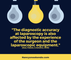
“Laparoscopic identification of superficial endometriosis implants represents a challenge for the gynecologic surgeon. Endometriosis lesions may present in a wide spectrum of appearance according to a “lifecycle” of the implants. The lesions can be flat or vesicular. They can have any combination of color typically red, back/brown and white. Active “red” lesions, large endometriomas, deep infiltrating nodules, and typical “powder-burn” lesions are easier to identify than “white” old fibrotic lesions. The endometriotic implants are hypervascular. The diagnostic accuracy at laparoscopy is also affected by the experience of the surgeon and the laparoscopic equipment [16].”
- Redwine, D. B., & Yocom, L. B. (1990). A serial section study of visually normal pelvic peritoneum in patients with endometriosis. Fertility and sterility, 54(4), 648-651. Retrieved from https://www.fertstert.org/article/S0015-0282(16)53823-0/abstract
“Visually normal peritoneum does not harbor a high prevalence of invisible microscopic endometriosis.”
Locations:
Links:
- Surgery Videos:
- “Posterior endometriosis” –– Dr Andrea Vidali MD: https://m.facebook.com/story.php?story_fbid=1499501720113159&id=151983631531648&ref=m_notif¬if_t=group_comment_reply&hc_location=ufi
- “Simple Endometriosis Excision”- Dr. Cindy Mosbrucker: https://vimeo.com/channels/453744
- “Endometriosis Excision by L.I.T.E. technique”- Dr. James Kondrup: https://www.youtube.com/watch?v=wK3yjUQQKOw&t=2&fbclid=IwAR1mKxl-1GeMXKbKdY29SVQ9RW2PAdYbcRkjX5Q0i1vAijOvjbJjE8mOR6c&has_verified=1
- “About endometriosis”: http://endometriosis.org/endometriosis/
- See “Weird places endometriosis has been found”
Studies:
- Samreen, J. N., Bookwalter, C. A., Burnett, T. L., Feldman, M., Sheedy, S. P., Menias, C., … & Kabashi, A. (2019). MRI of endometriosis: A comprehensive review. Applied Radiology, 48(5), 6-12. Retrieved from https://appliedradiology.com/communities/MR-Community/mri-of-endometriosis-a-comprehensive-review
“In the pelvis, endometriosis commonly involves the peritoneum (anterior and posterior cul-de-sacs, pelvic side walls and ovarian fossa), uterosacral ligaments, ovaries, fallopian tubes, and uterus. Other structures less commonly involved include the rectovaginal septum, rectum, sigmoid colon, appendix, ureters, and bladder. Additional extra-pelvic endometriosis is uncommon but can involve the diaphragm, cecum, small and large bowel, abdominal wall, and other abdominal organs.4 Other areas to assess include the inguinal canals, sciatic and sacral nerves, peri-hepatic region and pleura (Figure 1). There are three forms of intraperitoneal pelvic endometriosis. These include superficial peritoneal lesions (ie, noninvasive implants), endometriomas (ie, cystic endometriosis), and deep (or solid and/or cystic) infiltrating endometriosis (DIE).”
- Chopra, K., Dutta, D., & Jain, K. (2018). Surgical Management of Endometriosis-A Mini Review. Clin Case Rep Open Access, 1(2), 111. Retrieved from https://www.yumedtext.com/files/publish/published-pdf–6-CCR-111.pdf
“The different forms of endometriotic lesions are superficial peritoneal implants, endometriomas and deep infiltrative endometriosis involving rectovaginal septum. Peritoneal implants are most commonly seen in the uterosacral ligaments, pouch of douglas, ovarian fossae and pelvic side walls. Less frequently, bladder and bowel are involved and rarely upper abdomen as well. Gastrointestinal involvement is seen is 3%-37% cases, with severe endometriosis affecting uterosacral ligaments, rectovaginal septum, rectosigmoid colon and appendix [7,8]. Genitourinary endometriosis is seen in 1%-2% cases with ureter involvement in 0.1%-0.2% cases, with majority affecting distal ureter. Typically, the left ureter is involved and endometriosis leads to extrinsic compression of the ureter leading to irreversible damage to renal function. This, thus warrants timely diagnosis and management.”
- Charatsi, D., Koukoura, O., Ntavela, I. G., Chintziou, F., Gkorila, G., Tsagkoulis, M., … & Daponte, A. (2018). Gastrointestinal and urinary tract endometriosis: a review on the commonest locations of extrapelvic endometriosis. Advances in medicine, 2018. Retrieved from https://www.hindawi.com/journals/amed/2018/3461209/
“The gastrointestinal tract is the most common location of extrapelvic endometriosis with the urinary system being the second one. However, since sigmoid colon, rectum, and bladder are pelvic organs, extragenital pelvic endometriosis may be a more suitable definition for endometriotic implants related to these organs than extrapelvic endometriosis. The sigmoid colon is the most commonly involved, followed by the rectum, ileum, appendix, and caecum. Most lesions are confined in the serosal layer; however, deeper lesion can alter bowel function and cause symptoms. Bladder and ureteral involvement are the most common sites concerning the urinary system. Unfortunately, ureteral endometriosis is often asymptomatic leading to silent obstructive uropathy and renal failure. Surgical excision of the endometriotic tissue is the ideal treatment for all types of extrapelvic endometriosis. Adjunctive treatment might be useful in selected cases.”